
Advancements in Medical Image Analysis: A Metric-driven review of AI, ML, and DL Methods
DOI:
https://doi.org/10.26438/ijcse/v13i10.1423Keywords:
Medical Image Analysis (MIA), Machine Learning (ML), Artificial Intelligence (AI), Deep Learning (DL), Convolutional Neural Networks (CNN)Abstract
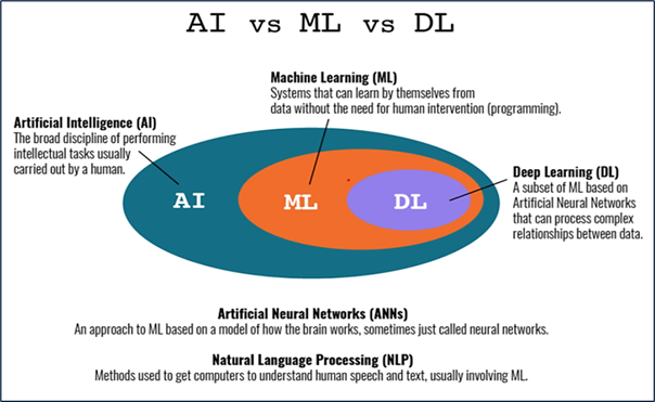
The domain of medical image analysis (MIA) within Machine Learning (ML), Artificial Intelligence (AI), and Deep Learning (DL), the significance of employing advanced methods cannot be overstated. Several methods have attained better outcomes in several fields, making it especially noteworthy for MIA in healthcare. The combination of these fields with MIA gives real-time analysis of prior and different databases, comprehensive insights that are important to improve healthcare results, and operational efficacy in the industry. This analysis article of existing literature considers a thorough examination of the most current ML, DL, and AI methods designed to identify the complexities faced in medical healthcare, specifically focusing on the utilization of DL, ML, and AI methods in MIA. The main contribution of this paper is how AI, ML and DL is used in medical field for early disease detection, drug discovery, and robotic-assisted surgeries. Comparative Analysis Based on the Different models and Algorithm is properly defined in this paper. It analysed the different methods, such as convolutional neural networks(CNNs), Support Vector Machine (SVM), Logistic Regression (LR), Decision Tree (DT), etc. This paper review the existing result was analysed, by other authors with a high accuracy achieved with the CNN method of 94%, XGBoost achieved the maximum accuracy of 91%, and Logistic Regression an accuracy of 79% and compared with the existing models.
References
[1] H. K. Huang, “Handbook of Medical Imaging,” Physics Today, USA, Vol.1–3, pp.57–57, 2001. doi: 10.1063/1.1420518.
[2] D. L. Rubin, N. H. Shah, and N. F. Noy, “Biomedical Ontologies: A Functional Perspective,” Briefings in Bioinformatics, Vol.9, No.1, pp.75–90, 2007. doi: 10.1093/bib/bbm059.
[3] P. Kaur and R. K. Singh, “A Review on Optimization Techniques for Medical Image Analysis,” Concurrency and Computation: Practice and Experience, Vol.35, No.1, 2022. doi: 10.1002/cpe.7443.
[4] J. Wang, “Recent Optimization Methods and Techniques for Medical Image Analysis,” Preprints, 2023. doi: 10.20944/preprints202309.2137.v1.
[5] M. Puttagunta and S. Ravi, “Medical Image Analysis Based on Deep Learning Approach,” Multimedia Tools and Applications, Springer, pp.1–15, 2021. doi: 10.1007/s11042-021-10707-4.
[6] G. Litjens et al., “A Survey on Deep Learning in Medical Image Analysis,” Medical Image Analysis, Vol.42, pp.60–88, 2017. doi: 10.1016/j.media.2017.07.005.
[7] P. Rajpurkar et al., “Deep Learning for Chest Radiograph Diagnosis: A Retrospective Comparison of the CheXNeXt Algorithm to Practicing Radiologists,” PLoS Medicine, Vol.15, No.11, pp.1–17, 2018. doi: 10.1371/journal.pmed.1002686.
[8] F. Liao, M. Liang, Z. Li, X. Hu, and S. Song, “Evaluate the Malignancy of Pulmonary Nodules Using the 3-D Deep Leaky Noisy-OR Network,” IEEE Transactions on Neural Networks and Learning Systems, Vol.30, No.11, pp.3484–3495, 2019. doi: 10.1109/TNNLS.2019.2892409.
[9] C. T. Sari and C. Gunduz-Demir, “Unsupervised Feature Extraction via Deep Learning for Histopathological Classification of Colon Tissue Images,” IEEE Transactions on Medical Imaging, Vol.38, No.5, pp.1139–1149, 2019. doi: 10.1109/TMI.2018.2879369.
[10] M. Anjum et al., “Congruent Feature Selection Method to Improve the Efficacy of Machine Learning-Based Classification in Medical Image Processing,” Computer Modeling in Engineering & Sciences, Vol.142, No.1, pp.357–384, 2024. doi: 10.32604/cmes.2024.057889.
[11] J. S. H. Baxter and R. Eagleson, “Exploring the Values Underlying Machine Learning Research in Medical Image Analysis,” Medical Image Analysis, Vol.102, pp.103494, 2025. doi:10.1016/j.media.2025.103494.
[12] A.Heena et al., “Machine Learning Based Biomedical Image Processing for Echocardiographic Images,” Multimedia Tools and Applications, Springer, 2022. doi: 10.1007/s11042-022-13516-5.
[13] G. Varoquaux and V. Cheplygina, “Machine Learning for Medical Imaging: Methodological Failures and Recommendations for the Future,” NPJ Digital Medicine, Vol.5, No.1, pp.1–8, 2022. doi: 10.1038/s41746-022-00592-y.
[14] K. Desai, “Diagnosis of Medical Images Using Convolutional Neural Networks,” Deleted Journal, Vol.20, No.6s, pp.2371–2376, 2024. doi: 10.52783/jes.3220.
[15] G. J. Trivedi and R. Sanghvi, “Medical Image Fusion Using CNN with Automated Pooling,” Indian Journal of Science and Technology, Vol.15, No.42, pp.2267–2274, 2022. doi: 10.17485/ijst/v15i42.1812.
[16] P. Kalyani et al., “Medical Image Processing from Large Datasets Using Deep Learning,” Proc. of 3rd Int. Conf. on Advances in Computing, Communication Control and Networking (ICAC3N), IEEE, India, pp.400–404, 2021. doi: 10.1109/icac3n53548.2021.9725513.
[17] S. S. Yadav and S. M. Jadhav, “Deep Convolutional Neural Network Based Medical Image Classification for Disease Diagnosis,” Journal of Big Data, Vol.6, No.1, 2019. doi: 10.1186/s40537-019-0276-2.
[18] M. Khalifa and M. Albadawy, “AI in Diagnostic Imaging: Revolutionising Accuracy and Efficiency,” Computer Methods and Programs in Biomedicine Update, Vol.5, pp.100146, 2024. doi: 10.1016/j.cmpbup.2024.100146.
[19] L. Pinto-Coelho, “How Artificial Intelligence is Shaping Medical Imaging Technology: A Survey of Innovations and Applications,” Bioengineering, Vol.10, No.12, pp.1435, 2023. doi: 10.3390/bioengineering10121435.
[20] A.Azizi, M. Azizi, and M. Nasri, “Artificial Intelligence Techniques in Medical Imaging: A Systematic Review,” International Journal of Online and Biomedical Engineering (IJOE), Vol.19, No.17, pp.66–97, 2023. doi: 10.3991/ijoe.v19i17.42431.
[21] H. J. Yoon et al., “Medical Image Analysis Using Artificial Intelligence,” Korean Society of Medical Physics, Vol.30, No.2, pp.49–58, 2019. doi: 10.14316/pmp.2019.30.2.49.
[22] N. F. Kingsley and A. C. Izuchukwu, “Optimization of Medical Image Analysis Models for Effective Disease Diagnosis through Data Augmentation Techniques,” Journal of Infectious Diseases and Patient Care, 2025.
[23] Y. Wu, M. Owais, R. Kateb, and A. Chaddad, “Deep Modeling and Optimization of Medical Image Classification,” arXiv preprint, arXiv:2505.23040, 2025.
[24] J. Wang, “Recent Optimization Methods and Techniques for Medical Image Analysis,” Preprints, 2023. doi: 10.20944/preprints202309.2137.v1.
[25] X. Li, Z. Wu, F. Zhang, and D. Qu, “Robust DC Optimization and Its Application in Medical Image Processing,” Technology and Health Care, Vol.29, No.2, pp.393–405, 2021. doi: 10.3233/thc-202656.
[26] L. P. Zhuhadar and M. D. Lytras, “The Application of AutoML Techniques in Diabetes Diagnosis: Current Approaches, Performance, and Future Directions,” Sustainability, Vol.15, No.18, pp.13484–13484, 2023. doi: 10.3390/su151813484.
[27] J. Zhuang et al., “Deep kNN for Medical Image Classification,” Proc. of MICCAI 2020: Medical Image Computing and Computer-Assisted Intervention, Springer, pp.127–136, 2020. doi: 10.1007/978-3-030-59710-8_13.
Downloads
Published
How to Cite
Issue
Section
License

This work is licensed under a Creative Commons Attribution 4.0 International License.
Authors contributing to this journal agree to publish their articles under the Creative Commons Attribution 4.0 International License, allowing third parties to share their work (copy, distribute, transmit) and to adapt it, under the condition that the authors are given credit and that in the event of reuse or distribution, the terms of this license are made clear.

